

Indian Journal of Plastic Surgery
Analysis of facial bone fractures: An 11-year study of 2,094 patients
Kun Hwang¹,², Sun Hye You¹
¹Department of Plastic Surgery and ²Center for Advanced Medical Education by BK21 Project, Inha University College of Medicine, Incheon, Korea
Address for correspondence: Dr. Kun Hwang, Department of Plastic Surgery and Center for Advanced Medical Education by BK21 Project, Inha University College of Medicine, 7-206 Sinheung-dong, Jung-gu, Incheon - 400-711, Korea. E-mail: jokerhg@inha.ac.kr
Free full text on www.ijps.org · DOI: 10.4103/0970-0358.63959 · Indian J Plast Surg January-June 2010 Vol 43 Issue 1
ABSTRACT
Purpose: The medical records of these patients were reviewed and analysed to determine the clinical characteristics and treatment of facial bone fractures.
Patients and Methods: This is a retrospective study of 2,094 patients with facial bone fractures from various accidents that were treated at the Inha University Hospital from 1996 to 2007.
Results: The most common age group was the third decade of life (29%). Males were more common than females (3.98:1). The most common aetiology was violent assault or nonviolent traumatic injury (49.4%). The most common isolated fracture site was the nasal bone (37.7%), followed by the mandible (30%), orbital bones (7.6%), zygoma (5.7%), maxilla (1.3%) and the frontal bone (0.3%). The largest group with complex fractures included the inferior region of the orbital floor and zygomaticomaxilla (14%). Closed reduction was performed in 46.3% of the cases while 39.7% of the cases required open reduction. For open reductions, the most commonly used soft-tissue approach was the intraoral approach (32.3%). The complication rate was 6.4% and the most common complication was hypoesthesia (68.4%) followed by diplopia (25.6%).
Conclusion: Long-term collection of epidemiological data regarding facial fractures and concomitant injuries is important for the evaluation of existing preventive measures and useful in the development of new methods of injury prevention and treatment.
KEY WORDS
Facial bones; facial fractures; injury prevention; treatment
INTRODUCTION
Facial injuries occur in a significant proportion of trauma patients requiring prompt diagnosis of fractures and soft tissue injuries, with possible emergency interventions.[1] Each year, increasing numbers of patients are admitted to the hospital with facial trauma.[1] There are many studies in the literature that have analysed the demographic factors associated with facial trauma according to various criteria.[2-4] The epidemiology of facial fractures varies with regard to injury type, severity and cause, depending on the population studied.[5] The differences in the populations with regard to the causes of facial fractures may be the result of differences in culture and a variety of risk factors. However, the reports on patients studied, use the severity of the injury as the major selection criteria for epidemiological investigation.[6]
An understanding of the cause, severity and temporal distribution of facial trauma can aid in establishing clinical and research priorities for effective treatment and prevention of these injuries.[6] Continuous long-term collection of data regarding the epidemiology of facial …
Indian J Plast Surg January-June 2010 Vol 43 Issue 1 · 42
Table 2: Distribution according to age and gender
| Age | Male | Female | Cases (%) |
|---|---|---|---|
| 0–10 | 64 | 31 | 95 (4.5) |
| 11–20 | 396 | 70 | 466 (22.3) |
| 21–30 | 188 | 120 | 608 (29.0) |
| 31–40 | 349 | 90 | 439 (21.0) |
| 41–50 | 222 | 73 | 295 (14.1) |
| 51–60 | 94 | 21 | 115 (5.5) |
| >60 | 60 | 16 | 76 (3.6) |
| Total cases (%) | 1,673 (79.9) | 421 (20.1) | 2,094 (100) |
Table 3: Annual incidence
| Year | Cases | % |
|---|---|---|
| 1996. 2 ~ | 64 | 3.1 |
| 1997 | 163 | 7.8 |
| 1998 | 155 | 7.4 |
| 1999 | 156 | 7.5 |
| 2000 | 177 | 8.5 |
| 2001 | 242 | 11.6 |
| 2002 | 219 | 10.5 |
| 2003 | 214 | 10.2 |
| 2004 | 184 | 8.8 |
| 2005 | 217 | 10.4 |
| 2006 | 271 | 12.9 |
| ~ 2007. 4 | 32 | 1.5 |
| Total | 2,094 | 100 |
… (n = 159, 7.6%) and others (n= 5 8, 2.8%) [Table 5]. The most common sports associated with injury was soccer (38.1%).
In 60 (2.9%) of the 2,094 patients, the facial fractures were associated with other injuries [Table 6]. Head and neck injuries were the most common isolated injuries associated with facial fractures (13.3%) [Table 6]. Among the patients with injuries to the head and neck area, most had intra cranial injuries with altered levels of consciousness, cervical spine injuries, or optic nerve injury. Some patients (1.3%) had more than one type of associated injury, including other bone fractures [Table 7]. The most common isolated fracture associated with facial fractures was a skull fracture (22.2%) [Table 7]. In 359 (17.1%) of the 2,094 patients, the facial fractures were associated with other soft tissue injuries [Table 8]. The associated injuries were most commonly soft tissue injuries of the face and neck (89.4%) [Table 8].
Classification of facial bone fractures and treatment
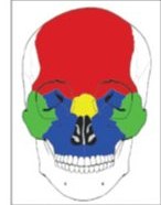
The great majority of cases were isolated injuries (n = 1720, 82.1%) [Table 1]. Nasal bone fractures were the most common (n = 790, 37.7%), followed by mandible fractures (n = 618, 30%) [Table 1, Figure 1]. Tripod fractures were the most common type of complex injuries (n = 130, 6.2%) [Table 1]. For complex injuries, the inferior region had the highest frequency of fractures (n = 293, 14%) [Table 1, Figure 2].
A closed reduction procedure was performed in 46.3% of the cases, open reduction in 39.7%, closed and open reduction in 6.5% and no surgical intervention in 7.4% [Table 9]. The closed reduction procedure was most commonly performed for nasal bone fractures (93%). Most of the other facial bone fractures were treated by open reduction (39.7%) procedures. The facial bone reduction …
Figure 1
The skeletal region of isolated facial bone fractures: red, frontal bone (0.4%); yellow, orbital bone (9.2%); green, nasal bone (45.9%); blue, maxilla (1.6%); purple, zygoma (6.9%); orange, mandible (35.9%)
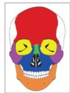
Figure 2
The skeletal region of complex facial bone fractures: red, superior (1.6%); yellow, medial (4.3%); green, lateral (1.1%); blue, inferior (78.3%)
Indian J Plast Surg January-June 2010 Vol 43 Issue 1 · 44