

The Journal of Craniofacial Surgery
Analysis of Nasal Bone Fractures; A Six-year Study of 503 Patients
Kun Hwang, MD, PhD, Sun Hye You, MD, Sun Goo Kim, MD, MS, Se Il Lee, MD, DMSc
Incheon, Korea
The aim of this study is to classify the nasal bone fractures based on computed tomography (CT) analysis and patterns of the nasal bone fractures, and review 503 cases treated between 1998–2004 at the Department of Plastic Surgery, Inha University Hospital, Incheon, South Korea.
The age, sex, etiology, associated injuries, pattern of fractures and treatments were reviewed and a radiographic study was analyzed. Plain simple radiographs of lateral and Waters view of the nasal bones combined with computed tomography scans were done. Nasal bone fractures were classified into six types: Type I) Simple without displacement; Type II) Simple with displacement/ without telescoping; IIA; Unilateral; IIAs) Unilateral with septal fracture; IIB) Bilateral; IIBs) Bilateral with septal fracture; Type III) Comminuted with telescoping or depression.
Diagnosis of nasal bone fractures were made positively by plain x-ray films in 82% of cases, negative finding was 9.5% and 8.5% of cases were suspicious of the fractures. Reliability of the plain film radiographs of the nasal bone fracture was 82% in this study. In the most of the fractured nasal bones (93%) the closed reduction was done, open reduction in 4% and no surgical intervention in 3%. Nasal reduction was carried out in average 6.5 days post the injury. The patterns of the nasal bones fractures classified by CT findings were type IIA (182 cases, 36%), IIBs (105 cases, 21%), IIB (90 cases, 18%), IIAs (66 cases, 13%), I (39 cases, 8%) and III (21 cases, 4.3%).
We think the CT is necessary for diagnosing nasal bone fracture because the reliability of the plain film was only 82%.
From the Department of Plastic Surgery, Inha University Hospital, Incheon, Korea. · Address correspondence and reprint requests to Kun Hwang, MD, PhD, Department of Plastic Surgery, College of Medicine, Inha University, 7-206 Sinheung-dong, Jung-gu, Incheon 400-711, Korea. · This work was supported by a grant from Inha University (INHA Research Grant). The authors thank Robert S. Chung, MD, FACS NB Plastic Surgical Institute, Newport Beach, CA, for editing this article. · THE JOURNAL OF CRANIOFACIAL SURGERY / VOLUME 17, NUMBER 2 March 2006 · Copyright © 2006 Mutaz B. Habal, MD. Unauthorized reproduction of this article is prohibited.
Key Words
Fractures, nasal bone
The nasal bone fracture is the most common type of facial bone fracture. The fractures have been described and classified by many authors. Stranc¹ classified the type of nasal fractures based on a pattern of impaction and level of injuries.
Kazanjian and Converse² reported a classification of nasal bone fractures and Murray³ also set a pathologic classification of nasal bone fractures.
However, no classification of fractured nasal bones has been appropriately applied to the clinical management. A detailed mechanism of the injury, a physical examination, and simple radiologic examination are essential to make a diagnosis of nasal bone fracture. An additional computed tomography (CT) image shows more accuracy of the nature of the fractured nasal bone. A CT analysis contributes to the nature of the fractures of nasal bone, associated injuries and decision of making plan and prognosis.
Many authors reported etiology and patterns of facial bone fractures in various areas,⁴,⁵ but not many of nasal bone fractures.
The aim of this study is to classify the nasal bone fractures based on CT analysis and patterns of the nasal bone fractures, and review 503 cases treated between 1998-2004 at the Department of Plastic Surgery, Inha University Hospital, Incheon, South Korea.
Clinical Material
Five hundred and three patients with nasal bone fractures were seen from January, 1998 to October, 2004 at the department of plastic and reconstructive surgery, Inha University Hospital, Incheon metropolitan city, South Korea.
The age, sex, etiology, associated injuries, pattern of fractures and treatments were reviewed and a radiographic study was analyzed. Simple radiographs of lateral and Waters view of the nasal bones combined with computed tomography scans were done.
261
Fig 1
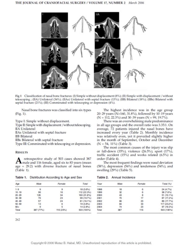
Classification of nasal bone fractures: (I) Simple without displacement (8%); (II) Simple with displacement / without telescoping ; (IIA) Unilateral (36%); (IIAs) Unilateral with septal fracture (13%); (IIB) Bilateral (18%); (IIBs) Bilateral with septal fracture (21%); (III) Comminuted with telescoping or depression (4%).
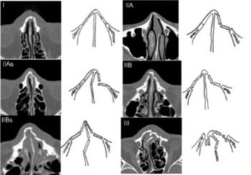
Nasal bone fractures was classified into six types (Fig. 1).
- Type I
Simple without displacement.
- Type II
Simple with displacement / without telescoping.
- IIA
Unilateral
- IIAs
Unilateral with septal fracture
- IIB
Bilateral
- IIBs
Bilateral with septal fracture
- Type III
Comminuted with telescoping or depression.
Results
A retrospective study of 503 cases showed 387 male and 116 female, aged six to 83 years (mean age = 29.2) with diverse fracture of nasal bones (Table 1).
The highest incidence was in the age group 20–29 years (N=160, 31.8%), followed by 10–19 years (N = 112, 22.3%) and 30–39 years (N = 99, 19.7%).
There was an overwhelming male predominance in all age groups and the overall ratio was 3.35:1. On average, 72 patients injured the nasal bones have increased every year (Table 2). Monthly incidence was relatively even, yet it prevailed slightly higher in the month of September, October and December (N = 54, 11%) (Table 3).
The most common causes of the injury was slip or fall-down (35%), violence (26.5%), sport (17%), traffic accident (15%) and works related (6.5%) in order (Table 4).
The most frequent findings were nasal deviation (58%), depression (56%) and tenderness (54%), and swelling (29%) (Table 5).
Table 1. Distribution According to Age and Sex
| Age | Male | Female | Total |
|---|---|---|---|
| 1-9 | 9 | 9 | 18 (3.6%) |
| 10-19 | 96 | 16 | 112 (22.3%) |
| 20-29 | 126 | 34 | 160 (31.8%) |
| 30-39 | 76 | 23 | 99 (19.7%) |
| 40-49 | 57 | 24 | 81 (16.1%) |
| 50-59 | 13 | 6 | 19 (3.8%) |
| 60+ | 9 | 5 | 14 (2.8%) |
| Total | 387 (77%) | 116 (23%) | 503 (100%) |
Table 2. Annual Incidence
| Year | Male | Female | Total (%) |
|---|---|---|---|
| 1998 | 18 | 6 | 24 (4.7%) |
| 1999 | 32 | 6 | 38 (7.6%) |
| 2000 | 43 | 8 | 51 (10.1%) |
| 2001 | 70 | 17 | 87 (17.2%) |
| 2002 | 66 | 23 | 89 (17.7%) |
| 2003 | 84 | 33 | 117 (23.2%) |
| 2004 | 74 | 23 | 97 (19.3%) |
| Total | 387 | 116 | 503 (100%) |
262